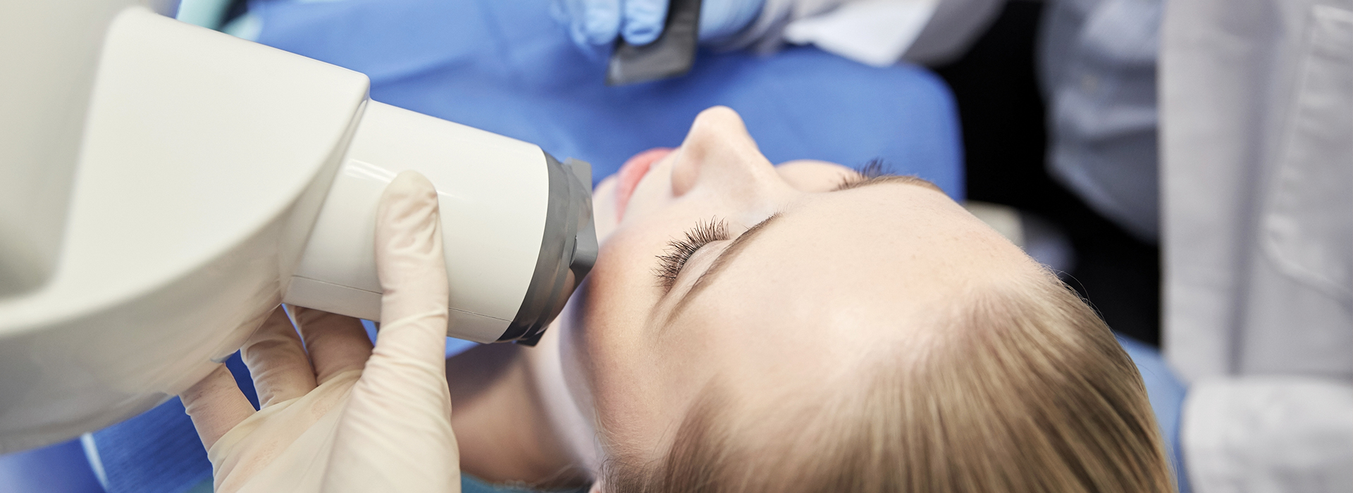

Digital radiography replaces traditional film with electronic sensors and computer imaging to capture detailed pictures of teeth, gums, and jawbone. For patients, the most noticeable differences are speed and clarity: images appear on a monitor within seconds, allowing your dental team to review them together during the appointment. Because the process integrates directly with electronic health records, images become a durable part of your chart that the clinical team can reference throughout your care.
Beyond convenience, digital radiography helps the practice provide more consistent, evidence-based treatment. High-resolution images reveal subtle changes that can be difficult to see on older film x-rays, supporting earlier detection of decay, fractures, and bone changes. That early visibility often translates into simpler, more conservative treatment options and better long-term outcomes for patients.
At Liberty Dental Care PC by Park One Dental, we use digital imaging as a routine component of diagnostic workflows so clinicians can make informed decisions quickly. Integrating digital radiography with other diagnostic tools helps our team deliver comprehensive evaluations with a focus on patient comfort and clinical accuracy.
One of the key advantages of digital radiography is that it generally requires less radiation than conventional film x-rays. Advanced sensors capture images more efficiently, which lowers the amount of exposure needed while still producing diagnostically useful detail. This reduction in radiation is particularly important for children, seniors, and patients who require frequent imaging as part of ongoing treatment.
Image quality is also markedly improved. Digital systems produce sharper contrast and finer resolution, and clinicians can enhance images with software tools—adjusting brightness, contrast, or zoom—without re-taking the x-ray. These capabilities make it easier to identify early-stage cavities, hairline fractures, and subtle bone changes that might otherwise be missed until they progress.
Because images are available immediately, there is no waiting for film to develop. That speed shortens appointments and reduces patient anxiety by allowing real-time explanation of findings. When necessary, images can be printed or exported for second opinions or collaboration with specialists, but most often they remain accessible within the secure digital record.
Digital radiography begins with a compact sensor placed in or near the mouth to capture x-ray photons that pass through dental structures. These sensors convert the incoming energy into a digital signal that a computer translates into a high-resolution image. There are a few sensor types in common use, but all share the same goal: precise image capture with minimal discomfort for the patient.
Once an image is acquired, specialized software processes and stores it in the patient’s electronic chart. The software provides measurement tools and image-enhancement features that clinicians use to examine fine details. For example, the team can measure the width of a root canal, assess bone levels around a tooth, or compare images over time to track healing and disease progression.
The workflow is designed for safety and efficiency. Sensors are covered with protective sleeves and disinfected according to infection-control protocols. Digital files are saved on secure systems and backed up regularly, ensuring that images remain accessible for future treatment planning or referrals without the risk of physical film deterioration.
Digital radiography supports nearly every aspect of modern dental care. For routine exams, bitewing images detect interproximal decay between teeth. Periapical views reveal root anatomy and signs of infection at the tooth tip, which is essential for endodontic assessment. Panoramic and cone-beam images (when indicated) provide a broader view of the jaws and airway for implant planning, surgical evaluation, and orthodontic assessment.
Because the images are readily available, the dental team can discuss findings with patients during the same visit, using the screen to point out areas of concern and explain recommended next steps. This visual approach helps patients understand their condition and participate in treatment decisions. Clinicians also rely on serial digital images to monitor healing after procedures such as implant placement or periodontal therapy.
Digital imaging aids interdisciplinary care as well. When collaboration with specialists is needed—whether for oral surgery, endodontics, or prosthodontics—digital files can be shared securely, enabling coordinated treatment planning without delay. That interoperability improves clinical outcomes and streamlines care for patients who require multiple services.
Patient safety is central to the way digital radiography is used in dental practices. In addition to lower radiation doses, strict protocols govern sensor handling, image acquisition settings, and protective barriers to maintain hygiene and minimize repeat exposures. Clinicians are trained to select the appropriate type and number of images for each clinical situation, avoiding unnecessary imaging while ensuring diagnostic adequacy.
Digital files are managed within secure electronic health record systems that provide organized storage, access controls, and routine backups. This approach eliminates the environmental hazards associated with film processing chemicals and reduces the physical storage burden of paper films. It also makes it easier to retrieve prior images for comparison, which is important for tracking changes in oral health over time.
Quality assurance remains an ongoing priority: sensors and software are calibrated regularly, image-acquisition protocols are reviewed, and staff receive continuing education so the team remains up to date with best practices. These measures help ensure that every image is useful for diagnosis and that patient care benefits from consistent, reproducible results.
In summary, digital radiography is a powerful, patient-friendly tool that enhances diagnostic clarity while improving office efficiency and reducing environmental impact. If you would like to learn more about how we use digital imaging in our practice or what to expect during your visit, please contact us for more information.
Digital radiography is a modern method for capturing dental x-ray images using electronic sensors and computer software instead of traditional film. The sensor records the image and transmits it instantly to a computer, where the image can be enhanced, stored, and reviewed. This digital workflow streamlines recordkeeping and supports more precise communication between dental team members.
Because images display immediately, dentists can review findings with patients in real time and adjust viewing settings to improve clarity. Digital radiography forms the foundation for many contemporary diagnostic tools, including image analysis and three-dimensional imaging systems used in advanced cases.
Unlike film x-rays, digital radiography captures images with sensors that convert x-ray energy into electronic signals, eliminating the need for chemical processing. Digital images can be brightened, contrasted, and magnified on a computer screen, which often makes it easier to identify small problems that might be missed on film. The turnaround time is also much faster since images appear instantly without development steps.
Digital files are easier to organize and share with other providers when coordination of care is needed, and they remove the environmental concerns associated with film processing chemicals. These workflow and image-quality advantages have made digital radiography the preferred standard in many dental offices.
Digital radiography is considered safe and is designed to use the lowest radiation dose necessary to produce a diagnostically useful image. Modern sensors are more sensitive than film, so they require less exposure to x-rays to capture a clear picture. Dental teams follow strict safety protocols, including shielding and appropriate exposure settings, to protect patients.
For most patients, routine digital dental x-rays present minimal risk and are an important diagnostic tool for monitoring oral health. Pregnant patients or those with specific medical concerns should inform the dentist so that exposure can be minimized and precautions can be taken as appropriate.
Digital radiography improves diagnostic accuracy by providing high-resolution images that can be enhanced and magnified to evaluate teeth, bone, and surrounding structures. These images help dentists detect cavities, bone loss, infections, and other conditions earlier than might be possible with a visual exam alone. Clear digital records also aid long-term monitoring and comparison over time.
Because images are immediately available on-screen, they support more efficient consultations and allow the dental team to explain findings directly to patients. The ability to share images electronically with specialists or laboratories also speeds up collaborative treatment planning and improves overall care coordination.
Digital sensors are more sensitive to x-rays than traditional film, so they require less radiation to produce a usable image. Advances in sensor design and imaging software further enhance image quality at lower doses, and modern x-ray units are calibrated to use minimal exposure for routine intraoral and extraoral images. When indicated, techniques such as collimation and appropriate exposure settings further limit the x-ray beam to the necessary area.
Additionally, digital systems often require fewer retakes because the captured image can be immediately evaluated and adjusted, reducing repeat exposures. Together, these factors contribute to a significant reduction in patient radiation when compared with older film-based workflows.
During a digital x-ray appointment, a small electronic sensor is placed inside the mouth or positioned externally depending on the type of image needed. The sensor is aligned with the x-ray source, and a brief exposure creates the image that is instantly transmitted to the computer. The entire capture process typically takes only a few seconds per image.
Preparation and positioning add a few minutes to ensure patient comfort and proper alignment, so a standard set of intraoral x-rays can usually be completed in a short appointment. Because images appear immediately, the dentist can review them with the patient without delay and request any additional views if clinically necessary.
Yes, one of the practical strengths of digital radiography is the ease of sharing files with specialists, referring providers, or other offices. Digital images can be exported in standard formats and securely transmitted via encrypted email, secure portals, or direct network transfer, which facilitates coordinated care. This capability reduces the need for patients to carry physical films and speeds up consultations and treatment planning.
When images are shared, dental teams typically include clinical notes to provide context and support clear communication. Patients should ask their provider about the office's procedures for secure transfer and storage of radiographic records if they have privacy concerns.
Dentists use digital radiography to reveal issues not visible on a clinical exam, such as interproximal cavities between teeth, root infections, impacted teeth, and bone loss from periodontal disease. Enhanced viewing tools allow clinicians to zoom in, adjust contrast, and measure structures accurately, improving the ability to diagnose early-stage problems. These detailed images guide decisions about restorations, root canal treatment, extractions, and periodontal therapy.
Digital radiographs also assist in assessing the fit and integrity of existing restorations and the position of developing teeth in younger patients. By combining digital images with clinical findings, dentists can develop more precise and individualized treatment plans for each patient.
Most patients experience minimal discomfort when having digital x-rays taken, as the sensors are small and noninvasive. Some people may find the sensor placement slightly awkward or sensitive, especially if they have a strong gag reflex or sore areas in the mouth, but the exposure itself is brief and painless. Dental staff are trained to position sensors carefully and to accommodate patient comfort during the procedure.
If you have concerns about discomfort or positioning, communicate them to the dental team before the x-ray is taken so adjustments can be made. Simple accommodations, such as taking breaks between images or using smaller sensors when appropriate, can make the experience more comfortable for many patients.
At Liberty Dental Care PC by Park One Dental, digital radiography is integrated into routine exams and diagnostic workflows to provide clear, immediate images that support accurate diagnosis and personalized treatment planning. These images help the doctors evaluate dental and periodontal health, monitor changes over time, and explain findings directly to patients using the office computer display. The practice also uses digital imaging to coordinate care with specialists when advanced treatment is recommended.
By relying on digital records, the office improves efficiency in charting and communications while reducing environmental waste associated with film processing. Patients benefit from quicker appointments, better visual explanations, and a modern approach to imaging that prioritizes safety and diagnostic precision.
Liberty Dental Care PC by Park One Dental
112-10 Liberty Avenue, Richmond Hill, NY 11419Park One Dental
1601 Jericho Turnpike, New Hyde Park, NY 11040 (516) 354-0033