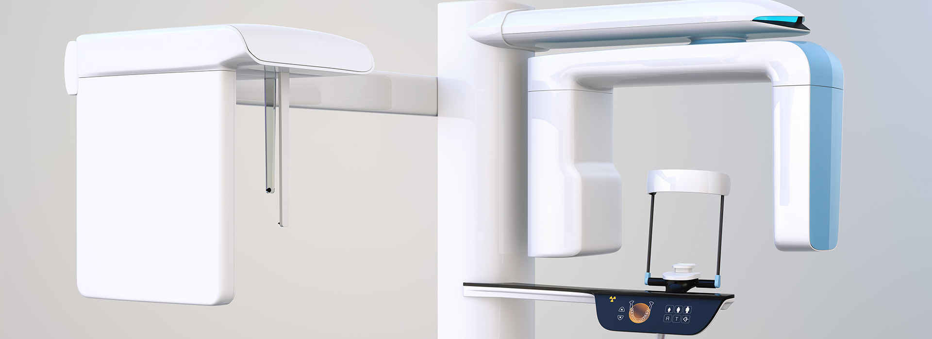

At Liberty Dental Care PC by Park One Dental, we prioritize accurate diagnosis and thoughtful treatment planning. One of the ways we deliver that standard of care is through cone-beam computed tomography (CBCT), a form of three-dimensional imaging designed specifically for dental and maxillofacial applications. CBCT allows our clinicians to see anatomical detail that traditional two-dimensional radiographs can’t reveal, helping us tailor treatments to each patient’s unique needs.
Using advanced imaging responsibly helps the practice provide safer, more predictable care. When appropriate, CBCT scans are incorporated into evaluations to clarify complex situations, confirm clinical findings, and support confident decision-making across restorative, surgical, and specialty treatments.
CBCT is a specialized X-ray technology that captures a series of images from multiple angles and reconstructs them into a single, three-dimensional view of the teeth, jaws, and surrounding structures. Unlike conventional panoramic or bitewing films, CBCT provides depth and perspective, enabling clinicians to evaluate spatial relationships with precision. The resulting volume dataset can be rotated and examined slice by slice for a comprehensive assessment.
The scan itself is typically quick — only a matter of seconds to a minute for most fields of view — and is targeted to the area of interest. That targeted approach reduces unnecessary exposure while giving clinicians the detailed information they need. CBCT units used in dental offices are optimized for oral and maxillofacial imaging rather than full-body diagnostic scans, producing diagnostic-quality images suited to dental care.
Because the technology produces anatomically accurate, distortion-free images, it is particularly helpful when two-dimensional radiographs leave questions unanswered. CBCT is a diagnostic tool that augments, rather than replaces, traditional imaging and clinical examination; together they create a more complete picture of oral health.
One of the most significant benefits of CBCT is its ability to reveal hidden or complex anatomy that affects treatment choices. For example, the orientation of tooth roots, the location of vital structures such as nerves and sinuses, and the presence of pathology or bone defects are more readily identified in 3D. This clarity helps clinicians anticipate challenges and select the most appropriate approach for a given case.
In restorative and implant dentistry, CBCT aids in determining whether a site has sufficient bone for stable implant placement and in choosing the optimal implant size and position. In endodontics, the technology can expose missed canals, confirm the extent of root fractures, or help locate the source of persistent symptoms. For patients, that means fewer surprises during treatment and a higher likelihood of successful outcomes.
Because CBCT datasets are digital, they can be integrated with other treatment tools — for example, to design surgical guides or to combine 3D models with intraoral scans. This interoperability supports precision workflows and helps align clinical intent with predictable results.
CBCT has broad utility across many areas of dental care. We commonly use it for implant planning, ensuring implants are positioned to avoid critical anatomy and to take full advantage of available bone. It is also valuable for assessing impacted teeth, evaluating complex root anatomy for endodontic treatment, and characterizing suspicious lesions or cysts that need further attention.
The technology is useful for diagnosing temporomandibular joint (TMJ) conditions, assessing airway space in select cases, and mapping sinus anatomy when upper-jaw procedures are planned. Orthodontic planning can also benefit from the additional context CBCT provides when growth patterns, skeletal relationships, or tooth impactions are in question.
Every scan is prescribed with a clinical purpose in mind. Our clinicians consider the potential diagnostic benefit, the anticipated impact on treatment decisions, and each patient’s individual concerns when determining whether CBCT is the most appropriate imaging choice.
Patient safety is central to how we use CBCT. Modern dental CBCT units are engineered to capture the information necessary for dental diagnosis while minimizing radiation exposure compared to diagnostic medical CT scans. We follow established guidelines for imaging and adhere to principles of justification and optimization — that is, only performing scans when they are expected to materially influence care, and using the smallest field of view that will answer the clinical question.
The scanning process is simple and well tolerated by most patients. The machine’s open design reduces feelings of confinement, and the short acquisition time helps minimize motion and discomfort. Our team explains each step before imaging and positions patients carefully to ensure clear, usable images on the first attempt.
Images are reviewed promptly by our clinicians, who interpret the findings within the context of the clinical exam and patient history. When additional input is helpful, collaboration with specialists ensures that complex findings are addressed with a multidisciplinary perspective.
CBCT is a cornerstone of contemporary digital dentistry because it enables workflows that link diagnosis directly to execution. When planning implants or guided surgeries, CBCT data can be used to create precise surgical guides that translate virtual plans into accurate clinical placement. That integration reduces variability and supports more predictable healing and prosthetic outcomes.
In combination with intraoral scanners and CAD/CAM systems, three-dimensional imaging helps produce restorations and appliances designed around the patient’s actual anatomy. This coordination shortens treatment timelines and improves fit and function for many procedures, from crowns to complex rehabilitations.
Beyond planning, CBCT datasets serve as a baseline record of anatomy and treatment intent, which can be useful for monitoring healing and for future reference should new issues arise. By capturing a volumetric snapshot of the jaws and surrounding structures, the practice can make well-informed choices throughout a patient’s continuum of care.
In summary, CBCT offers targeted, three-dimensional insight that enhances diagnostic confidence and supports more precise, individualized treatment. When used judiciously and in combination with a thorough clinical exam, it becomes a powerful tool for improving outcomes across many areas of dentistry.
If you have questions about CBCT or whether three-dimensional imaging is recommended for your care, please contact us for more information. Our team is happy to explain how this technology might benefit your treatment and what to expect during the imaging process.
Cone-beam computed tomography, commonly called CBCT, is an advanced dental imaging technique that captures three-dimensional views of the teeth, jaws, and surrounding structures. The system rotates around the patient's head to collect a series of images that are reconstructed into a detailed 3D model. These volumetric images show bone, tooth roots, nerve canals, and sinus anatomy with much greater clarity than traditional two-dimensional X-rays. Clinicians use CBCT to visualize complex anatomy and plan care with a high degree of precision.
CBCT imaging is widely used in modern dentistry because it provides targeted diagnostic information while remaining efficient and comfortable for patients. The scan captures a complete view quickly, helping the team evaluate conditions that are difficult to assess on standard films. Because data are three-dimensional, clinicians can view anatomy from multiple angles and create cross-sectional slices for exact measurements. This capability supports safer, more predictable treatment planning across many dental specialties.
Traditional dental X-rays produce two-dimensional images that flatten complex anatomy into a single plane, while CBCT generates true three-dimensional datasets that reveal depth and spatial relationships. Standard films are excellent for routine exams and detecting cavities, but they can miss important details like the precise position of a nerve canal or the true extent of bone defects. CBCT slices can be reformatted at various angles to show cross-sections, panoramic views, and volumetric models that are not possible with conventional radiography. This additional information helps clinicians make more informed diagnoses and plan treatments with greater accuracy.
The field of view of a CBCT scan can be adjusted to focus on a single tooth, a jaw segment, or the entire maxillofacial region, which provides flexibility that traditional X-rays lack. Because the images are digital, they can be measured, annotated, and integrated with surgical guides or implant planning software. This interoperability improves communication between the dental team and any specialists involved in care. For many complex procedures, CBCT offers a diagnostic advantage that directly contributes to better outcomes.
CBCT uses ionizing radiation, but modern cone-beam systems employ focused beams and optimized settings to minimize exposure while delivering diagnostic-quality images. Imaging is performed under the principle of ALARA (as low as reasonably achievable), and clinicians select the smallest appropriate field of view and lowest acceptable dose to answer the clinical question. Protective measures such as lead aprons and thyroid collars are used when indicated, and scans are only recommended when the expected benefit outweighs the minimal risk. For most adults, a properly performed CBCT scan is considered safe and provides critical information that cannot be obtained with routine X-rays.
Before ordering a CBCT scan, your dentist will evaluate whether the 3D information is necessary for diagnosis or treatment planning and will discuss the rationale with you. If you have concerns about radiation, the dental team can explain why the scan is recommended and how exposure is controlled. Records are kept to avoid unnecessary repeat imaging, and alternative imaging options are considered when appropriate. Clear communication ensures patients understand both the value and safety measures associated with CBCT.
CBCT is valuable across many areas of dental care, including implant planning, evaluation of impacted teeth, assessment of jaw pathology, and complex endodontic cases. For implant dentistry, three-dimensional images allow the clinician to measure bone volume, identify critical anatomical landmarks such as the inferior alveolar nerve and sinus cavities, and plan implant position with surgical guides. In endodontics, CBCT can reveal root fractures, unusual canal anatomy, or periapical lesions that are not visible on two-dimensional films.
Additional uses include TMJ analysis, airway assessment for sleep-disordered breathing, evaluation of trauma or fractures, and surgical planning for extractions and orthognathic procedures. CBCT can also assist in orthodontic diagnosis by showing tooth position relative to surrounding bone. Because the technology provides detailed anatomic context, it supports more accurate diagnoses and tailored treatment strategies across multiple specialties.
Preparation for a CBCT scan is minimal and straightforward, which makes it convenient for most patients. You may be asked to remove metal jewelry, eyeglasses, hearing aids, or removable dental appliances that could interfere with image quality. If you have a history of recent imaging, bring those records to your appointment so the dentist can determine whether additional scans are necessary.
If you are pregnant or suspect you might be, inform the dental team before the scan so they can evaluate alternatives or postpone imaging when appropriate. Follow any specific instructions provided by the office, such as arriving a few minutes early to complete forms or to review your medical history. Otherwise, no special preparation, fasting, or medication changes are typically required for a CBCT study.
A CBCT examination is generally quick and well tolerated, with actual image acquisition often completed in less than a minute. The overall appointment may take a little longer to position the patient and review safety measures, but the process is usually completed within a single visit. During the scan you will stand or sit still while the machine rotates around your head, and staff will be nearby to ensure proper positioning and comfort.
The procedure is painless and noninvasive, with no injections or sedation required in most cases. You may be asked to bite on a stabilizing device or hold a still pose briefly to reduce motion artifacts. After the scan, images are reconstructed and reviewed by the dentist or specialist, who will explain the findings and how they inform your treatment plan.
CBCT datasets provide precise anatomical detail that clinicians use to measure distances, assess bone quality, and identify critical structures prior to treatment. These measurements support surgical guides for implant placement, allow for virtual implant planning, and help predict potential complications by visualizing the relationship between teeth, nerves, and sinuses. In complex cases, CBCT images can be shared with specialists, labs, or referral providers to coordinate care and ensure a consistent plan.
Beyond static assessment, CBCT data can be combined with digital impressions, CAD/CAM systems, and surgical planning software to create custom guides and restorations. This digital workflow enhances predictability and can reduce chair time during the actual procedure. By integrating three-dimensional imaging into planning, clinicians can communicate expected outcomes more clearly and tailor treatments to each patient's unique anatomy.
Yes, CBCT often reveals pathology or anatomical details that two-dimensional imaging can overlook due to overlapping structures and limited perspective. It can identify lesions in the jawbone, small fractures, root resorption, and the true extent of cysts or tumors with greater clarity than standard films. For endodontic evaluation, CBCT can show untreated canals, vertical root fractures, or periapical pathology that might be hidden on periapical radiographs.
While CBCT is a powerful diagnostic tool, it is used selectively when the additional information will influence care. The dentist interprets the scans in the context of clinical findings and may recommend further evaluation or referral when abnormal findings are detected. This targeted approach ensures that CBCT contributes to accurate diagnosis without unnecessary imaging.
CBCT can be used for pediatric patients when the clinical benefit outweighs the minimal radiation exposure, and protocols are adjusted to use the lowest possible dose suitable for the diagnostic task. Pediatric imaging requires careful consideration of field of view, exposure settings, and immobilization to reduce motion and dose. The dental team will weigh alternatives and choose age-appropriate imaging strategies to obtain the necessary diagnostic information while protecting the child's long-term health.
For pregnant patients, CBCT is typically avoided unless an urgent, clinically justified need exists that cannot be addressed by other means. If imaging is required during pregnancy, the team will take additional precautions and use the most conservative settings and shielding to protect the patient and fetus. Always inform the dental staff if you are pregnant or believe you may be pregnant so they can evaluate options and plan care accordingly.
Liberty Dental Care PC by Park One Dental incorporates advanced CBCT technology into a comprehensive, patient-centered practice to support precise diagnoses and predictable treatment outcomes. Our team emphasizes careful assessment, appropriate use of imaging, and clear communication so patients understand why a scan is recommended and how results inform care. By combining modern equipment with experienced clinicians, we aim to deliver safe, efficient imaging that adds value to your treatment plan.
When CBCT is indicated, we tailor the scan to the clinical question and follow established safety protocols to minimize exposure while maximizing diagnostic benefit. Patients can expect professional handling of their images, integration with digital treatment planning tools, and thoughtful follow-up to discuss findings and next steps. Our goal is to provide the information clinicians need to plan effective, individualized care for every patient.
Liberty Dental Care PC by Park One Dental
112-10 Liberty Avenue, Richmond Hill, NY 11419Park One Dental
1601 Jericho Turnpike, New Hyde Park, NY 11040 (516) 354-0033